
 EN
EN



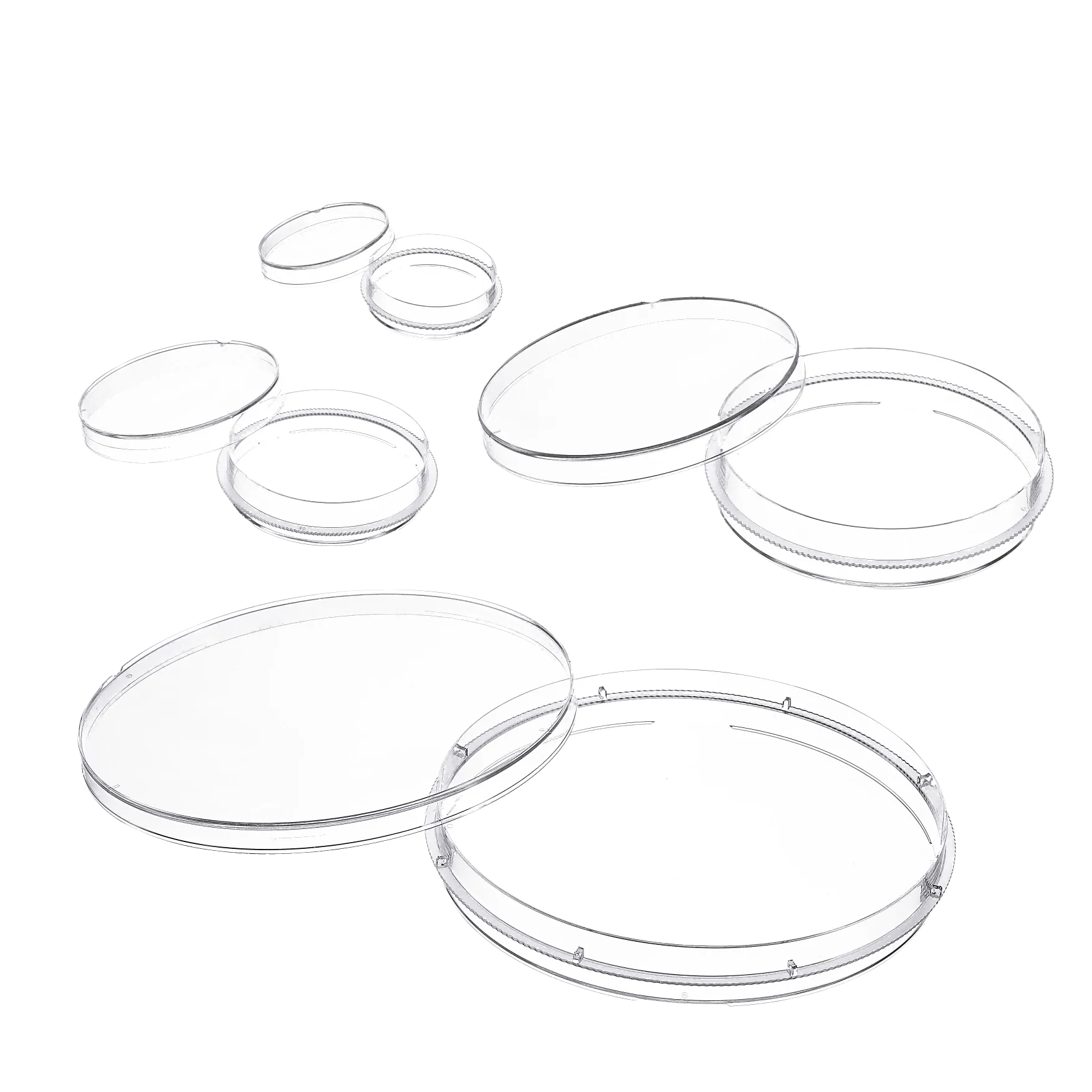
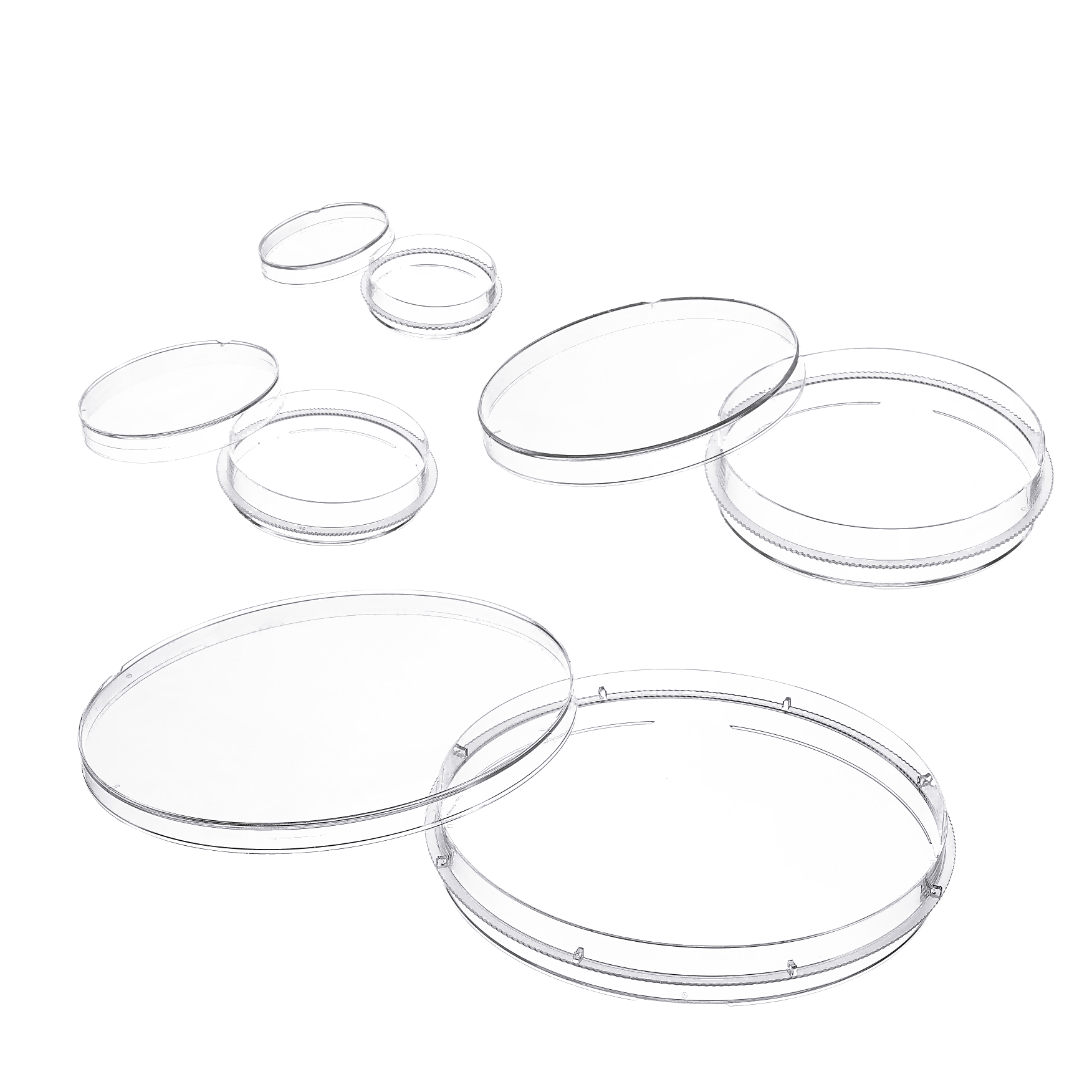
In modern biotechnology laboratories, few tools are as foundational and widely relied upon as cell culture dishes. These flat, circular, precision-engineered containers serve as the primary environment in which living cells are grown, maintained, and studied under controlled conditions. From pharmaceutical research to regenerative medicine, cell culture dishes have become indispensable components of the scientific workflow, enabling researchers to replicate complex biological processes outside of living organisms. Their design, surface treatment, and material composition are all carefully optimized to support cell adhesion, proliferation, and viability across a wide spectrum of applications.
Understanding the specific applications of cell culture dishes in biotechnology helps researchers select the right format for each experimental need and make informed decisions about laboratory consumables. This article explores the major use cases in which cell culture dishes play a critical role, from basic cell biology experiments to advanced therapeutic manufacturing. Whether you are setting up a new research lab or scaling up a bioproduction process, knowing where and how cell culture dishes are applied will guide your procurement and experimental design decisions effectively.
Cell-Based Research and Fundamental Biology Studies
Growing and Maintaining Adherent Cell Lines
One of the most common and essential applications of cell culture dishes in biotechnology is the routine maintenance of adherent cell lines. Adherent cells, which require a solid surface to attach and spread before they can divide, depend entirely on the treated polystyrene surface found in standard cell culture dishes. The surface is typically treated with a hydrophilic coating that promotes cell attachment, mimicking in vivo tissue environments closely enough to sustain normal cellular behavior.
Researchers routinely use cell culture dishes to passage cell lines at regular intervals, maintaining stocks of well-characterized cells for ongoing experiments. Popular cell lines such as HeLa, HEK293, and CHO cells are all grown and subcultured in cell culture dishes of various diameters, typically ranging from 35 mm to 150 mm. The larger dishes provide greater surface area for harvesting higher cell numbers, while smaller formats are ideal for experiments requiring precise, smaller-scale conditions.
Because the growth environment in cell culture dishes is highly controllable, researchers can regulate temperature, CO2 levels, and nutrient availability with precision. This level of control makes cell culture dishes the preferred vessel for long-term cell maintenance in both academic and industrial biotechnology settings.
Studying Cell Morphology and Behavior
Beyond simple cell maintenance, cell culture dishes are widely used to observe and study cell morphology, motility, and structural changes over time. Because cell culture dishes are manufactured from optically clear materials, they are compatible with transmitted-light microscopy, phase contrast imaging, and fluorescence microscopy. This transparency is critical when monitoring changes in cell shape, cytoskeletal organization, or the formation of cell clusters.
Researchers studying cell migration, division cycles, or stress responses regularly use cell culture dishes as observation platforms. Time-lapse imaging experiments, for example, involve growing cells in cell culture dishes placed directly onto an incubated microscope stage, allowing live monitoring of dynamic cellular events without disturbing the culture environment. The standardized flat geometry of cell culture dishes ensures consistent focal distances across the dish surface, improving image quality and reproducibility.
These fundamental biology applications in cell culture dishes provide the raw data that underpins drug discovery pipelines, toxicology assessments, and mechanistic studies of disease at the cellular level.
Drug Discovery and Pharmacological Testing
High-Throughput Compound Screening
In pharmaceutical biotechnology, cell culture dishes serve as the initial testing ground for new drug candidates. Early-stage compound libraries are screened against disease-relevant cell lines grown in cell culture dishes to identify molecules with potential therapeutic activity. The ability to culture large numbers of cells in standardized conditions makes cell culture dishes an efficient platform for performing dose-response studies, cytotoxicity assays, and receptor binding experiments.
Researchers often use cell culture dishes in combination with plate readers, automated liquid handling systems, and imaging platforms to accelerate the pace of data collection. Large-diameter cell culture dishes allow for the parallel preparation of multiple treatment groups, reducing the number of individual experiments needed to generate statistically meaningful results. This scalability is one of the primary reasons cell culture dishes remain central to preclinical drug discovery workflows despite the rise of more complex three-dimensional culture models.
The reproducibility offered by high-quality cell culture dishes is particularly important during the hit-to-lead and lead optimization phases of drug development, where small variations in cell behavior can obscure meaningful biological signals and delay candidate selection.
Toxicity and Safety Assessment
Toxicological evaluation of chemical compounds, environmental agents, and novel therapeutic molecules frequently relies on cells grown in cell culture dishes. In vitro toxicity testing performed in cell culture dishes has become a core component of safety profiling in both pharmaceutical and chemical industries, providing a cost-effective and ethically acceptable alternative or complement to animal testing.
Cell viability assays, oxidative stress measurements, and apoptosis detection are all routinely conducted in cell culture dishes. Hepatocyte cultures, for example, are grown in cell culture dishes to evaluate drug-induced liver injury — one of the leading causes of late-stage drug development failure. Primary human cells sourced from different tissue types can be seeded into cell culture dishes and exposed to test compounds to generate organ-specific toxicity profiles relevant to clinical safety predictions.
This widespread use of cell culture dishes in toxicological research reflects their reliability, consistency, and compatibility with a broad range of detection methods, from colorimetric assays to flow cytometry and Western blotting protocols run on cell lysates prepared directly from the dish.
Virology and Infectious Disease Research
Virus Propagation and Titration
Cell culture dishes have been central to virology research for decades. Viruses cannot replicate independently, so they require living host cells grown in environments such as cell culture dishes to complete their replication cycles. Virologists seed permissive cell monolayers into cell culture dishes, infect them with a viral inoculum, and then harvest viral particles from the resulting supernatant once an appropriate replication period has passed.
The plaque assay, a classical method for determining viral titer, is performed directly in cell culture dishes. A diluted virus suspension is applied to a confluent cell monolayer in a cell culture dish, and after incubation, plaques — clear zones of cell death caused by viral spread — are counted to calculate the concentration of infectious viral particles. This technique, which has changed relatively little since its development in the mid-twentieth century, continues to be the gold standard in infectious disease research and vaccine production quality control.
During the development of vaccines and antiviral therapies, cell culture dishes serve as the workhorse for both amplifying viral stocks and evaluating the inhibitory effects of candidate compounds on viral replication kinetics.
Pathogen-Host Interaction Studies
Beyond simple virus propagation, cell culture dishes are used to study the molecular mechanisms by which pathogens invade, subvert, and destroy host cells. Bacterial pathogens, intracellular parasites, and prions are all studied using host cell monolayers maintained in cell culture dishes. These experiments allow researchers to dissect the virulence factors of pathogens, understand host immune responses, and identify new intervention targets.
Fluorescent labeling, immunofluorescence imaging, and confocal microscopy are commonly applied to infected cells grown in cell culture dishes to visualize the intracellular journey of pathogens and monitor the cellular damage they cause. The flat, optically clear geometry of cell culture dishes is particularly advantageous for high-resolution imaging of infection events at the cellular level.
The COVID-19 pandemic accelerated global investment in infectious disease research infrastructure, and cell culture dishes were at the heart of early efforts to culture SARS-CoV-2, study its replication in human airway cells, and screen antiviral compound libraries for therapeutic leads.
Stem Cell Biology and Regenerative Medicine
Stem Cell Expansion and Differentiation
Stem cell biology represents one of the most demanding and rapidly advancing areas in which cell culture dishes are applied. Both pluripotent stem cells — including embryonic stem cells and induced pluripotent stem cells — and adult tissue stem cells require specialized culture conditions that cell culture dishes must support. For many stem cell types, the surface chemistry of the dish is modified with extracellular matrix proteins such as Matrigel, fibronectin, or laminin to promote attachment and maintain an undifferentiated state.
Large-scale expansion of stem cells for therapeutic manufacturing relies on consistent, reproducible performance from cell culture dishes across hundreds or even thousands of individual culture vessels. Any variation in surface treatment, material quality, or dimensional tolerances can introduce variability into cell expansion efficiency, which in turn affects downstream differentiation protocols and the yield of therapeutically relevant cell populations.
Directed differentiation protocols, in which stem cells are guided toward specific cell lineages such as cardiomyocytes, hepatocytes, or neural progenitors, are also typically initiated and conducted in cell culture dishes. The dish serves as the controlled stage on which carefully timed additions of growth factors and small molecules guide cell fate decisions at each stage of the differentiation timeline.
Tissue Engineering and Organoid Development
While traditional cell culture dishes support two-dimensional monolayer cultures, recent advances in biotechnology have expanded their role into three-dimensional culture applications. Low-attachment cell culture dishes with non-adherent surfaces are used to promote the self-assembly of cells into spheroids and organoids — miniaturized, three-dimensional tissue models that more accurately mimic the architecture and function of human organs than standard flat cultures.
Tumor spheroids grown in non-adherent cell culture dishes are used to model solid tumors in three dimensions, capturing the hypoxic core, proliferating rim, and necrotic center that characterize real tumor masses. These more physiologically relevant models are increasingly used in cancer drug development to predict in vivo drug responses more accurately than standard monolayer assays performed in regular cell culture dishes.
For tissue engineering applications aimed at producing transplantable tissues, cell culture dishes serve as the initial seeding platform before cells are transferred to scaffolds or bioreactor systems. The preparation, characterization, and quality control of cell populations used in these regenerative medicine workflows all depend heavily on cell culture dishes as the primary culture vessel.
Bioproduction and Recombinant Protein Manufacturing
Transient Transfection and Gene Expression
In biotechnology manufacturing, cell culture dishes are extensively used during the development phase of recombinant protein and viral vector production processes. Transient transfection experiments, in which plasmid DNA encoding a target protein is introduced into mammalian cells, are routinely performed in cell culture dishes at research scale before processes are transferred to bioreactors for larger production runs.
The ability to control cell density, transfection reagent concentration, and DNA dose precisely within the defined surface area of cell culture dishes makes them ideal for optimizing gene expression conditions. Researchers can evaluate multiple promoter constructs, transfection reagents, and incubation conditions in parallel using cell culture dishes of consistent format and quality, generating the data needed to define an optimal manufacturing process before committing to expensive scale-up efforts.
Cell culture dishes used in these applications must meet strict quality standards, including low background fluorescence for imaging-based readouts and minimal extractable chemical content that could interfere with biological assays or downstream analytical measurements of protein expression levels.
Cell Line Development and Clone Selection
During cell line development for biologics manufacturing, researchers isolate and evaluate individual clones derived from transfected cells. Cell culture dishes are used at multiple stages of this process, from plating transfected populations at low density to identify single-cell colonies, to expanding selected high-producing clones for further characterization. The flat, open surface of cell culture dishes makes it straightforward to visually identify and manually pick individual colonies under a microscope for isolation and expansion.
Stable cell line development workflows rely on cell culture dishes as the primary vessel for initial colony formation under selection pressure, typically achieved by including selection antibiotics in the culture medium. Over successive passages in cell culture dishes, non-expressing cells die off while stable integrants continue to proliferate, allowing researchers to identify the highest-producing clones for scale-up into production bioreactors.
The quality, sterility, and consistency of cell culture dishes used in these stages directly influence the success rate of cell line development programs, making supplier selection and product quality assurance critical considerations for biopharmaceutical development teams.
FAQ
What sizes of cell culture dishes are most commonly used in biotechnology laboratories?
Cell culture dishes are available in several standard diameters, with 35 mm, 60 mm, 100 mm, and 150 mm being the most widely used formats. Smaller dishes are preferred for experiments requiring limited cell numbers or precious reagents, while larger dishes are used when high cell yields are needed for downstream applications such as protein extraction, RNA isolation, or large-scale compound treatment studies. The choice of dish size is typically guided by the experimental scale, available incubator space, and the volume of culture medium required to maintain cell health.
How do treated cell culture dishes differ from untreated ones?
Treated cell culture dishes undergo a surface modification process — commonly corona discharge or plasma treatment — that increases the hydrophilicity of the polystyrene surface, promoting protein adsorption and cell adhesion. Treated dishes are appropriate for most adherent cell types that naturally attach to extracellular matrix components. Untreated or low-attachment cell culture dishes, by contrast, have surfaces that resist protein binding and cell adhesion, making them suitable for suspension cultures, spheroid formation, and organoid development where uncontrolled cell attachment would disrupt the culture system.
Can cell culture dishes be reused after sterilization?
Standard cell culture dishes are manufactured as single-use disposable labware and are not designed for reuse. Autoclaving or chemical sterilization can alter the surface chemistry of cell culture dishes, compromising cell adhesion, changing optical clarity, and potentially introducing chemical contaminants that affect cell viability or experimental results. For research requiring reusable culture surfaces, specialized glass-bottom dishes or dedicated reusable culture vessels with validated sterilization protocols are available, though single-use cell culture dishes remain the industry standard in most laboratory settings due to their convenience and assured sterility.
What materials are cell culture dishes typically made from?
The vast majority of cell culture dishes used in biotechnology are manufactured from medical-grade polystyrene, a material chosen for its optical clarity, dimensional stability, ease of surface modification, and low cost of production. Some specialized cell culture dishes are made from cyclic olefin copolymer materials that offer superior optical properties for advanced imaging applications. Glass-bottom cell culture dishes combine a glass coverslip base with a plastic dish wall, providing the optical performance of glass with the familiar format of standard cell culture dishes, making them particularly popular in confocal and super-resolution microscopy workflows.